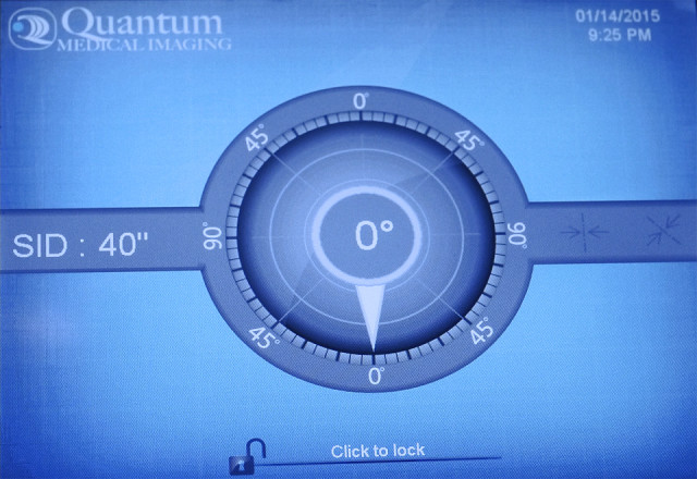
SID.
Meet SID! He's the monitor of our x-ray tube and his full name is "Source Image receptor Distance". What this means in plain English is that most x-rays images are captured 40 inches from the patient, but sometimes that distance is increased to 72 inches. X-Ray images are obtained by casting the "shadows" of soft tissue or bony anatomy onto an image receptor. In the old days that receptor was a sheet of film. Now it is most often a digital phosphor. The casting of those shadows can be manipulated by how close or far the x-ray source is from the patient's anatomy, and the level of detail necessary for an image to be of diagnostic value can thus be optimized. Showing the spaces between bones (or joints) requires that the machine be angled to match the patient's anatomy. That's what the quadrant is for. On older machines the adjustments were set with analog dials. Now, of course, it's a digital display. And yet the basic technology is pretty-much unchanged since the end of the 19th century.
Comments
Sign in or get an account to comment.


