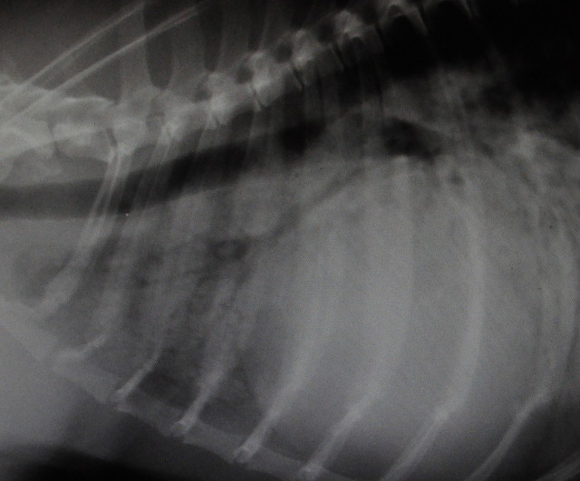
Air-bronchograms
This is a portion of the radiograph of a very breathless dog.
She was conscious for this examination - anaesthesia would've killed her - this means there is some movement blur on the film.
The blip shows the front part of the chest. The lung tissues, or the alveoli, which should be virtually black on a radiograph (because they are normally filled with air) are consolidated in this radiograph appearing off-white, which means that the bronchi (tubes of the lungs) show up as dark branching columns. I don't think I've ever seen as clear an example of this in all my years as a vet.
Lung consolidation and air-bronchograms are seen in a variety of very serious conditions including pneumonia, pulmonary oedema, lung tumours etc. This poor dog has pulmonary oedema and pneumonia; a very nasty double whammy, but as of this evening seems to be improving on treatment. The heart shadow on this radiograph is enlarged, somewhat globular, but difficult to examine completely. She has heart disease which is contributing to the lung disease.
Note:
Most radiographs of animals must be taken under deep sedation or anaesthesia for health and safety of staff (in the UK , at least, the rules are very clear on this). But, exceptionally, if a patient is very very fragile we are permitted to radiograph animals who are conscious, provided the staff are never exposed to the primary beam of the X Rays and that they wear lead gowns and other protective shielding.
Comments
Sign in or get an account to comment.


